Jane Oner
Jane Oner
  • Blog
  • About
  • Contact

Solution For Bursitis In Ball Of Foot

8/26/2015

 
Overview

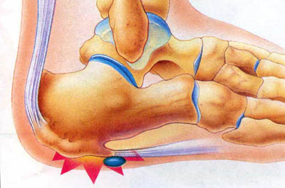
Between the heel bone and the plantar fascia is a small fluid filled sac that is called the plantar (bottom of) calcaneal (heel bone) bursa. The plantar calcaneal bursa?s job is to allow the plantar fascia to glide easily over the heel bone without irritation. If this becomes inflamed and irritated a plantar calcaneal bursitis occurs which can be extremely painful and disabling forcing the athlete to stop training.

Causes

Inflammation of the bursa causes synovial cells to multiply and thereby increases collagen formation and fluid production. A more permeable capillary membrane allows entrance of high protein fluid. The bursal lining may be replaced by granulation tissue followed by fibrous tissue. The bursa becomes filled with fluid, which is often rich in fibrin, and the fluid can become hemorrhagic. One study suggests that this process may be mediated by cytokines, metalloproteases, and cyclooxygenases.

Symptoms

Below is a list of common signs and symptoms of retrocalcaneal bursitis. Recognizing and treating symptoms early can prevent retrocalcaneal bursitis from becoming chronic. Swelling. The retrocalcaneal bursa is located behind the Achilles tendon, just above where the tendon attaches to the heel bone. When the bursa is inflamed it will cause visible soft tissue swelling near the top of the heel bone. It is worth noting that bursitis of the retroachilles bursa, which is located between the Achilles tendon and skin, can manifest slightly differently: swelling may be more distinct, appearing as a hard lump behind the heel. Retroachilles bursitis is also more likely than retrocalcaneal bursitis to cause the skin at the back of the heel to turn red.

Diagnosis

Bursitis is usually diagnosed after a careful physical examination and a full review of your medical history. If you garden and spend a lot of time on your knees, or if you have rheumatoid arthritis, tell your doctor, this information can be very helpful. During the physical exam, he or she will press on different spots around the joint that hurts. The goal is to locate the specific bursa that is causing the problem. The doctor will also test your range of motion in the affected joint. Other tests usually aren?t required to diagnose bursitis, but your doctor may suggest an MRI, X-ray or ultrasound to rule out other potential causes of pain.

Non Surgical Treatment

Treatments should involve decreasing swelling, relieving pain and stress on the Achilles, correcting any biomechanical dysfunction (over-pronation or flat feet), treating scar tissue, and then restoring strength and movement. If you are performing an activity that could cause further trauma to the bursa, it is recommended that you protect the area with padding and/or proper footwear to prevent further irritation or damage.

Surgical Treatment

Bursectomy is a surgical procedure used to remove an inflamed or infected bursa, which is a fluid-filled sac that reduces friction between tissues of the body. Because retrocalcaneal bursitis can cause chronic inflammation, pain and discomfort, bursectomy may be used as a treatment for the condition when it is persistent and cannot be relived with other treatments. During this procedure, a surgeon makes small incisions so that a camera may be inserted into the joint. This camera is called an arthroscope. Another small incision is made so that surgical instruments can be inserted to remove the inflamed bursa.

Hammer Toe Caused By Joint Inflammation

6/26/2015

 
Hammer ToeOverview

A Hammer toes or contracted toe is a deformity of the proximal interphalangeal joint of the second, third, or fourth toe causing it to be permanently bent, resembling a hammer. Mallet toe is a similar condition affecting the distal interphalangeal joint.

Causes

But what causes the imbalance of the tendons and muscles in the first place so that they begin to pull and bend the joint? A bad fitting shoe could be the cause but it usually isn?t the primary cause. Many people are genetically predisposed to hammertoe, and the condition begins to progress more quickly when they wear shoes that fit poorly, for example pointy toes, high heels, or shoes that are too short. Hammertoe may also be caused by damage to the joint as a hammertoe result of trauma.

HammertoeSymptoms

Some people never have troubles with hammer toes. In fact, some people don't even know they have them. They can become uncomfortable, especially while wearing shoes. Many people who develop symptoms with hammer toes will develop corns, blisters and pain on the top of the toe, where it rubs against the shoe or between the toes, where it rubs against the adjacent toe. You can also develop calluses on the balls of the feet, as well as cramping, aching and an overall fatigue in the foot and leg.

Diagnosis

A hammertoe is usually diagnosed with a physical inspection of your toe. Imaging tests, such as X-rays, may be ordered if you have had a bone, muscle, or ligament injury in your toe.

Non Surgical Treatment

Hammertoes that are not painful (asymptomatic) and still flexible may not require treatment. In mild cases, open-toed, low-heeled, or wider shoes and foam or moleskin pads can provide symptomatic relief by reducing pressure. Taping (strapping) the affected toe can help to reduce deformity and pain. Physical therapy to instruct patients in exercises that passively stretch tight structures and strengthen weak foot intrinsic muscles is also helpful with mild cases. Periodic trimming (debridement) of corns (clavi, helomata) by a podiatrist can provide temporary relief. Corticosteroid injections are often very effective in reducing pain.

Surgical Treatment

Hammer toe can be corrected by surgery if conservative measures fail. Usually, surgery is done on an outpatient basis with a local anesthetic. The actual procedure will depend on the type and extent of the deformity. After the surgery, there may be some stiffness, swelling and redness and the toe may be slightly longer or shorter than before. You will be able to walk, but should not plan any long hikes while the toe heals, and should keep your foot elevated as much as possible.

Hammer Toe Relief

6/26/2015

 
HammertoeOverview

A hammertoes is commonly mistaken as any type of toe deformity. The terms claw toe, or mallet toe, although technically different than a hammer toe, are commonly referred as such. The toe may be flexible with movement at the joints, or it may be rigid, especially if it has been present for a long time. With a true hammertoe the deformity exists at the proximal interphalangeal joint only.

Causes

Footwear is actually the leading cause of this type of toe deformity so much so that people sometimes require hammer toe surgery to undo some of the damage. The most common problem is wearing shoes that are too short, too narrow or too tight. These shoes constricts the feet and force the toes into a Hammer toes bend position. Women are more at risk especially due to high heels. Footwear isn?t the only problem, poor foot posture can lead to muscle and even bone imbalances. This asymmetry can cause excessive strain on the toes either by forcing the toe into unnatural positions. Arthritis can also play a factor in the development of hammer toe, especially if the toe joint is stiff and incapable of a full range of motion.

Hammer ToeSymptoms

Patients with hammer toe(s) may develop pain on the top of the toe(s), tip of the toe, and/or on the ball of the foot. Excessive pressure from shoes may result in the formation of a hardened portion of skin (corn or callus) on the knuckle and/or ball of the foot. Some people may not recognize that they have a hammer toe, rather they identity the excess skin build-up of a corn.The toe(s) may become irritated, red, warm, and/or swollen. The pain may be dull and mild or severe and sharp. Pain is often made worse by shoes, especially shoes that crowd the toes. While some hammer toes may result in significant pain, others may not be painful at all. Painful toes can prevent you from wearing stylish shoes.

Diagnosis

Although hammertoes are readily apparent, to arrive at a diagnosis the foot and ankle surgeon will obtain a thorough history of your symptoms and examine your foot. During the physical examination, the doctor may attempt to reproduce your symptoms by manipulating your foot and will study the contractures of the toes. In addition, the foot and ankle surgeon may take x-rays to determine the degree of the deformities and assess any changes that may have occurred.

Non Surgical Treatment

Your podiatrist may recommend one or more of these treatments to manage your hammer toes. Wear shoes with roomy toe boxes which don?t force your toes together. Exercises to stretch and strengthen muscles in the toes. Over the counter toe straps, cushions, and corn pads. Custom orthotic inserts. Toe caps or toe slings. In severe cases, surgery to release the muscles in the affected toes.

Surgical Treatment

If you have a severe case of hammer toe or if the affected toe is no longer flexible, you may need surgery to straighten your toe joint. Surgery requires only a local anesthetic (numbing medicine for the affected area) and is usually an outpatient procedure. This means you don?t have to stay in the hospital for the surgery.

HammertoePrevention

It?s important to understand that preventing hammertoe can sometimes be difficult, since most symptoms do not appear until the condition is well developed. Nonetheless, here are some tips to help you prevent hammertoe. Do not wear shoes that are too narrow or short. Check your children?s shoe size often to ensure that their shoes still fit correctly. Wear comfortable shoes that fit you properly. Remember that your feet widen and lengthen with age.

Bunions Treatment Exercises

6/7/2015

 
Overview
Bunions Callous A bunion is a firm, painful bump that forms over a bony bulge at the base of the big toe. In most cases, the big toe joint also is enlarged and has degenerative arthritis. The toe also may be pushed toward the second toe (hallux valgus). Bunions tend to be inherited, but they also are common in the following groups. Women who wear high heels. People who wear shoes that are too narrow or too pointed. People with flatfeet. All of these situations force the big toe to drift toward the little toes, and this can cause bunions to form.

Causes
Essentially, bunions are caused by a disruption of the normal interworking of the bones, muscles, ligaments and tendons that comprise your feet, often from wearing shoes that squeeze the toes or place too much weight-bearing stress on them. However, it should be pointed out that other causes or factors in the development of bunions can include flat feet or low arches in the feet, some forms of arthritis, problems with foot mechanics, foot injuries and neuromuscular disorders such as cerebral palsy. Arthritis in the MTP joint, for example, can degrade the cartilage that protects it, and other problems may cause ligaments to become loose. Pronation, walking in a way that your foot rolls inwards, increases your risk for developing bunions.

Symptoms
The dominant symptom of a bunion is a big bulging bump on the inside of the base of the big toe. Other symptoms include swelling, soreness and redness around the big toe joint, a tough callus at the bottom of the big toe and persistent or intermittent pain.

Diagnosis
The doctor considers a bunion as a possible diagnosis when noting the symptoms described above. The anatomy of the foot, including joint and foot function, is assessed during the examination. Radiographs (X-ray films) of the foot can be helpful to determine the integrity of the joints of the foot and to screen for underlying conditions, such as arthritis or gout. X-ray films are an excellent method of calculating the alignment of the toes when taken in a standing position.

Non Surgical Treatment
If overpronation is diagnosed early enough, the mechanics of the feet can be adjusted using a prescription orthotic. If orthotics are worn consistently, many major foot deformities can be avoided such as bunions. Early detection is of paramount importance. When a bunion progresses and cannot be controlled by an orthotic, surgical correction may be a consideration. Many advances in bunion correction allow for surgical intervention to make healing and return to normal activities much easier than use of traditional bunion surgery. Bunion Pain

Surgical Treatment
One of the more popular proximal metatarsal osteotomies that is performed is called the Myerson/Ludloff procedure. This operation is performed for more advanced deformity. Screws are inserted into the metatarsal to hold the bone cut secure and speed up bone healing. Walking is permitted in a surgical shoe following surgery. The shoe is worn approximately 5 weeks.

Prevention
Because bunions develop slowly, taking care of your feet during childhood and early adulthood can pay off later in life. Keep track of the shape of your feet as they develop over time, especially if foot problems run in your family. Exercising your feet can strengthen them. Learn to pick up small objects, like a pencil or pebble, with your toes. Wear shoes that fit properly and don't cramp or pinch your toes. Women should avoid shoes with very high heels or pointed toes.

What Actually Will Cause Feet To Over Pronate

6/5/2015

 
Overview

Pronation describes a slight inward rolling motion the foot makes during a normal walking or running stride. The foot (and ankle) roles slightly inward to accommodate movement. Some people, however, over-pronate and roll more than normal. With over-pronation, the arch of the foot flattens and causes excessive stress and pressure on the soft tissues of the foot. Over-pronation is more common in those with flat feet, and can lead to foot aches and pain, such as plantar fasciitis, Shin Splints and Knee Pain.Over Pronation

Causes

Unless there is a severe, acute injury, overpronation develops as a gradual biomechanical distortion. Several factors contribute to developing overpronation, including tibialis posterior weakness, ligament weakness, excess weight, pes planus (flat foot), genu valgum (knock knees), subtalar eversion, or other biomechanical distortions in the foot or ankle. Tibialis posterior weakness is one of the primary factors leading to overpronation. Pronation primarily is controlled by the architecture of the foot and eccentric activation of the tibialis posterior. If the tibialis posterior is weak, the muscle cannot adequately slow the natural pronation cycle.

Symptoms

Overpronation may have secondary effects on the lower legs, such as increased rotation of the tibia, which may result in lower leg or knee problems. Overpronation is usually associated with many overuse injuries in running including medial tibial stress syndrome, or shin splints, and knee pain Individuals with injuries typically have pronation movement that is about two to four degrees greater than that of those with no injuries. Between 40% and 50% of runners who overpronate do not have overuse injuries. This suggests that although pronation may have an effect on certain injuries, it is not the only factor influencing their development.

Diagnosis

People who overpronate have flat feet or collapsed arches. You can tell whether you overpronate by wetting your feet and standing on a dry, flat surface. If your footprint looks complete, you probably overpronate. Another way to determine whether you have this condition is to simply look at your feet when you stand. If there is no arch on the innermost part of your sole, and it touches the floor, you likely overpronate. The only way to truly know for sure, however, is to be properly diagnosed by a foot and ankle specialist.Over-Pronation

Non Surgical Treatment

Fortunately, there are simple things you can do to cure and correct your overpronation issues. Certain exercises help. Pull your toes back using a rolled up towel. Roll your feet over a golf or tennis ball for a minute. And do calf raises by standing up and lifting up on your toes. These all help reposition the foot and strengthen the muscles and tendons necessary for proper support. Beyond that, simple adjustments to footwear will help immensely.

Surgical Treatment

Subtalar Arthroereisis. The ankle and hindfoot bones/midfoot bones around the joint are fused, locking the bones in place and preventing all joint motion. This may also be done in combination with fusion at other joints. This is a very aggressive option usually reserved for extreme cases where no joint flexibility is present and/or the patient has severe arthritic changes in the joint.

Physiotherapy For Severs Disease

5/16/2015

 
Overview

Sever?s disease is a common cause of heel pain, particularly in the young and physically active. Just before puberty the calf bones typically grow faster than the surrounding soft tissue, which means the Achilles tendon is pulled uncomfortably tight. This can lead to an injured heel. Treatment includes relative rest, modifying activities and teaching the young person how to manage the condition when a flare-up happens. Sever?s disease is self-limiting and rarely causes long-term problems.

Causes

Sever?s disease is directly related to overuse of the bone and tendons in the heel. This can come from playing sports or anything that involves a lot of heel movement. It can be associated with starting a new sport, or the start of a new season. Children who are going through adolescence are also at risk of getting it because the heel bone grows quicker than the leg. Too much weight bearing on the heel can also cause it, as can excessive traction since the bones and tendons are still developing. It occurs more commonly in children who over-pronate, and involves both heels in more than half of patients.

Symptoms

A few signs and symptoms point to Sever?s disease, which may affect one or both heels. These include pain at the heel or around the Achilles tendon, Heel pain during physical exercise, especially activities that require running or jumping, worsening of pain after exercise, a tender swelling or bulge on the heel that is sore to touch, calf muscle stiffness first thing in the morning, limping, a tendency to tiptoe.

Diagnosis

Sever?s disease can be diagnosed based on the symptoms your child has. Your child?s doctor will conduct a physical examination by squeezing different parts of your child?s foot to see if they cause any pain. An X-ray may be used to rule out other problems, such as a broken bone or fracture.

Non Surgical Treatment

Depending on the diagnosis and the severity of the pain, there a number of treatment options available. Rest, reduce activity, your child should reduce or stop any activity that causes pain, such as sports and running. This can be a difficult option, as children are normally quite willful in pursuit of their favorite pastimes. Over the counter anti-inflammatory drugs, such as ibuprofen (found in Nurofen), to help reduce pain and inflammation. Make certain your child does stretching exercises before play. This will often help reduce the stress on the fascia and relieve heel pain in your child. Orthotic insoles. Orthotics made for children will help support the foot properly, and help prevent over-pronation or improper gait by supporting your child?s foot into a proper biomechanical position. Do not hesitate to schedule an appointment with a podiatrist, should your child?s heel and foot pain persist.

Prevention

To reduce the risk of heel pain or sore heels from Sever?s Disease. Only wear properly fitting shoes. A lace up shoe with a firm heel counter. Stretch calf and foot before exercising or playing sports. Properly taping the foot provides excellent protection and immediate pain relief. Wear shoe inserts or an over-the-counter orthotic. If the problem persists, consult your foot doctor.

Working With Pain In The Arches

4/16/2015

 
Overview

Most people with flat feet have a condition that is referred to as a flexible flat foot. This is caused by excessive flexibility within the joints and ligaments of their foot and is often hereditary. This results in an over pronated (rolled in) position and is exaggerated by mechanical misalignment of the foot. This can be a very destructive position for the foot to function in and can cause a wide variety of foot, leg and lower back conditions. The appearance of flat feet may vary from foot to foot. Generally there is a collapse of the medial (inside/middle) long arch of the foot. This may occur mildly where the arch height drops slightly or can be a large movement that results in the entire length of the arch resting on the ground. The twisting and misalignment of the foot that occurs with arch collapse can cause excessive wear and tear of the foot joints and eventually pain.

Arch Pain

Causes

Spending a lot of time on your feet. Especially when you are not used to doing so. For example you may have started a new job such as waiting tables where you are on your feet all day and wake up the next day with sore feet. This is a sign of damage and over time could lead to plantar fasciitis. Being Over-Weight. Never an easy topic to discuss but in simple terms, the heavier you are, the greater the burden on your feet. There are times when you're walking when your entire body weight is borne on one leg and therefore one foot, placing great strain on the plantar fascia. Wearing shoes with poor arch support or cushioning. A tight Achilles tendon. This is the big tendon at the bottom of your calf muscles above your heel. If this is excessively tight this can affect your ability to flex your ankle and make you more likely to damage your plantar fascia. Suddenly changing your exercise routine. Using running as an example if you suddenly run many more miles than your are used to or change to a new running surface e.g. grass to tarmac - these factors can put excessive strain on the plantar fascia and lead to plantar fasciitis. All of these risk factors ultimately lead to a specific change in foot structure. The term given is over-pronation and this basically describes rolling in of the foot and lowering of the arches. It is this change that excessively elongates the plantar fascia which can lead to plantar fasciitis.

Symptoms

Persistant pain and selling under the ball of the foot and extending towards the toes (most commonly the 2nd). Some swelling may be disable on the top of the foot along with redness. Often a sensation of 'walking on the bones for the foot' will be described, and there is a positive Lachman's test. Often a tear will result in the toes splaying (daylight sign) and clawing.

Diagnosis

The doctor will examine your feet for foot flexibility and range of motion and feel for any tenderness or bony abnormalities. Depending on the results of this physical examination, foot X-rays may be recommended. X-rays are always performed in a young child with rigid flatfeet and in an adult with acquired flatfeet due to trauma.

Non Surgical Treatment

There are many treatments for fasciitis. The most common initial treatment provided by the family doctor are anti-inflammatory medications. They may take the edge off the pain, but they don't often resolve the condition fully. Steroid injections, which deliver the medication directly to the most painful area, are usually more effective. Rest, ice, weight loss, taping, strapping, immobilization, physiotherapy, massage, stretching, heel cushions, acupuncture, night splints and extra-corporeal shock wave therapy all help some patients.

Arch Pain

Surgical Treatment

If you have pain that has not been responsive to other treatments, there is a new non-surgical treatment that was recently approved by the FDA. ESWT (extracorporeal shockwave therapy) uses strong electrohydraulic acoustic (sound) energy that triggers the body?s natural repair mechanism. This treatment method is safe, effective and requires a very short recovery period compared to older surgical techniques.

Prevention

To prevent arch pain, it is important to build up slowly to your exercise routine while wearing arch supports inside training shoes. By undertaking these simple measures you can prevent the discomfort of arch pain which can otherwise linger for many months. While you allow the foot to recover, it will help to undertake low impact exercises (such as swimming or water aerobics).

Stretching Exercises

Strengthening exercises. Below are two simple strength exercises to help condition the muscles, tendons and joints around the foot and ankle. Plantar Rolling. Place a small tin can or tennis ball under the arch of the affected foot. Slowly move the foot back and forth allowing the tin can or tennis ball to roll around under the arch. This activity will help to stretch, strengthen and massage the affected area. Toe Walking. Stand upright in bare feet and rise up onto the toes and front of the foot. Balance in this position and walk forward in slow, small steps. Maintain an upright, balanced posture, staying as high as possible with each step. Complete three sets of the exercise, with a short break in between sets, for a total of 20 meters.

What Will Be The Causes Of  Achilles Tendon Rupture

4/12/2015

 
Overview Achilles Tendinitis 
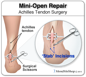
A rupture of the Achilles tendon is one of the more common types of tendon ruptures. As the largest tendon in the human body, the Achilles tendon is very important to one?s balance and stability. Patients who suffer an Achilles tendon rupture experience severe pain and often require Achilles tendon surgery to repair the damage. There are also less invasive methods of Achilles tendon treatment including, icing, anti-inflammatory medications, rehabilitation, taping or bracing, sports massages and more. Achilles tendon recovery is dependent on the health of the patient, and the severity of the injury. Causes Achilles tendon rupture occurs in people that engage in strenuous activity, who are usually sedentary and have weakened tendons, or in people who have had previous chronic injury to their Achilles tendons. Previous injury to the tendon can be caused by overuse, improper stretching habits, worn-out or improperly fitting shoes, or poor biomechanics (flat-feet). The risk of tendon rupture is also increased with the use of quinolone antibiotics (e.g. ciprofloxacin, Levaquin). Symptoms Although it's possible to have no signs or symptoms with an Achilles tendon rupture, most people experience pain, possibly severe, and swelling near your heel. An inability to bend your foot downward or "push off" the injured leg when you walk. An inability to stand up on your toes on the injured leg. A popping or snapping sound when the injury occurs. Seek medical advice immediately if you feel a pop or snap in your heel, especially if you can't walk properly afterward. Diagnosis A typical history as detailed above together with positive clinical examination usually will clinch the diagnosis. In an acute rupture, one can usually feel the gap in the tendon from the rupture. There may be swelling or bruising around the ankle and foot of the injured leg. With the patient lying on the tummy (prone position) with the knee flexed, the examiner should see the ankle and foot flex downwards (plantarward) when squeezing the calf muscles. If there is no movement in the ankle and foot on squeezing the calf muscle, this implies that the calf muscle is no longer attached to the heel bone due to a complete Achilles tendon rupture. Non Surgical Treatment Once the Achilles tendon is partially damaged, one should exercise great care. The risk of rupture is high and if pain is associated with walking, one should consult with an orthopedic surgeon or a sports physician. A complete rupture of the Achilles tendon is never treated at home. It is important to understand that there are no minerals, nutrients, or herbs to treat Achilles tendon injury and any delay just worsens the recovery. Achilles Tendon Surgical Treatment Surgical techniques for rupture repair are varied but usually involve reapproximation of the torn ends of the Achilles tendon, sometimes reinforced by the gastrocsoleus aponeurosis or plantaris tendon. Open reconstruction is undertaken using a medial longitudinal approach. Studies indicate that patients who undergo percutaneous, rather than an open, Achilles tendon rupture repair have a minimal rate of infection but a high rate of sural nerve entrapment (16.7% of treated cases). Prevention The following can significantly reduce the risk of Achilles tendon rupture. Adequate stretching and warming up prior to exercising. If playing a seasonal sport, undertake preparatory exercises to build strength and endurance before the sporting season commences. Maintain a healthy body weight. This will reduce the load on the tendon and muscles. Use footwear appropriate for the sport or exercise being undertaken. Exercise within fitness limits and follow a sensible exercise programme. Increase exercise gradually and avoid unfamiliar strenuous exercise. Gradual ?warm down? after exercising.

Achilles Tendon

3/29/2015

 
Overview
Achilles Tendinitis The Achilles tendon is the largest and most vulnerable tendon in the body. It joins the gastrocnemius (calf) and the soleus muscles of the lower leg to heel of the foot. The gastrocnemius muscle crosses the knee, the ankle, and the subtalar joints and can create stress and tension in the Achilles tendon . Tendons are strong, but not very flexible so they can only so far before they get inflammed and tear or rupture.

Causes
The Achilles tendon usually ruptures as a result of a sudden forceful contraction of the calf muscles. Activities such as jumping, lunging, or sprinting can cause undue stress on the Achilles tendon and cause it to rupture. Often there is a background of Achilles tendinitis. Direct trauma to the area, poor flexibility or weakness of the calf muscles or of the Achilles tendon and increasing age are some of the other factors that are associated with an Achilles tendon rupture.

Symptoms
Tendon strain or tendon inflammation (tendonitis) can occur from tendon injury or overuse and can lead to a rupture. Call your doctor if you have signs of minor tendon problems. Minor tenderness and possible swelling increases with activity. There is usually no specific event causing sudden pain and no obvious gap in the tendon. You can still walk or stand on your toes. Acute calf pain and swelling can indicate a tear or partial tear of the Achilles tendon where it meets the calf muscle. You may still be able to use that foot to walk, but you will need to see a specialist such as an orthopedic surgeon. Surgery is not usually done for partial tears. Sometimes special heel pads or orthotics in your shoes may help. Follow up with your doctor to check for tendonitis or strain before resuming activity, because both can increase the risk of tendon rupture. Any acute injury causing pain, swelling, and difficulty with weight-bearing activities such as standing and walking may indicate you have a tear in your Achilles tendon. Seek prompt medical attention from your doctor or emergency department. Do not delay! Early treatment results in better outcome. If you have any question or uncertainty, get it checked.

Diagnosis
It is usually possible to detect a complete rupture of the Achilles tendon on the history and examination. A gap may be felt in the tendon, usually 4-5cm above the heel bone. This is the normal site of injury and is called an intra-substance tear. The tear can occur higher up about 10cm above the insertion into the heel at the site where the muscles join the tendon, this is known as a musculo-tendinous tear. A special test will be performed which involves squeezing the calf. Normally if the Achilles tendon is intact this causes the foot to point downwards but if it is ruptured it causes no movement. To confirm the diagnosis and the exact site of the rupture it may be necessary to perform an Ultra-sound or MRI scan.

Non Surgical Treatment
Your doctor will advise you exactly when to start your home physical therapy program, what exercises to do, how much, and for how long to continue them. Alphabet Range of Motion exercises. Typically, the first exercise to be started (once out of a non-removable cast). While holding your knee and leg still (or cross your leg), you simply write the letters of the alphabet in an imaginary fashion while moving your foot and ankle (pretend that the tip of your toe is the tip of a pencil). Motion the capital letter A, then B, then C, all the way through Z. Do this exercise three times per day (or as your doctor advises). Freeze a paper cup with water, and then use the ice to massage the tendon area as deeply as tolerated. The massage helps to reduce the residual inflammation and helps to reduce the scarring and bulkiness of the tendon at the injury site. Do the ice massage for 15-20 minutes, three times per day (or as your doctor advises). Calf Strength exercises. This exercise is typically delayed and not used in the initial stages of rehabilitation, begin only when your doctor advises. This exercise is typically done while standing on just the foot of the injured side. Sometimes, the doctor will advise you to start with standing on both feet. Stand on a step with your forefoot on the step and your heel off the step. The heel and forefoot should be level (neither on your tip toes nor with your heel down). Lower your heel very slowly as low as it will go, then rise back up to the level starting position, again very slowly. This is not a fast exercise. Repeat the exercise as tolerated. The number of repetitions may be very limited at first. Progress the number of repetitions as tolerated. Do this exercise one to two times per day (or as your doctor advises). Achilles Tendon

Surgical Treatment
Thanks to a new surgical technique, operative procedures are often more beneficial.The operative treatment of Achilles tendon ruptures has significantly changed in recent years. The objective today is to connect the torn tendons using modern suture and possibly adhesive materials. Through small surgical incisions the ends of the torn tendon are surgically exposed, and sutures are used to tie the ends permanently together. Thus, the operated repaired tendon is again resilient within a reasonable time. The cast treatment and walking on crutches required in the past, is with this procedure usually not necessary. Instead, functional treatment following the surgery involves wearing a special boot, meaning that the patient can put weight onto the operated leg again within a few days after surgery. Physical therapy training will start immediately following the operation. The philosophy behind such an early functional treatment is that tissue adequately adapts to stress and thus accelerates the healing process. For the patient, the modern surgical treatment of an Achilles tendon rupture has the distinct advantage that no prolonged hospital stay is necessary. Hospital stay usually lasts only a few days. Using crutches, patients can return to work soon after the surgery.
Powered by Create your own unique website with customizable templates.